Overview
A dysplastic nevus, often called an atypical mole, is a mole that looks different from common moles. While most dysplastic nevi are benign (not cancerous), they can sometimes look like melanoma, a serious type of skin cancer, and people with multiple dysplastic nevi have a higher risk of developing melanoma.
These moles are thought to develop due to a combination of genetic factors and sun exposure. Anyone can develop a dysplastic nevus, but they are more common in individuals with a family history of melanoma or many moles, and often appear during puberty or young adulthood, though they can develop at any age. Dysplastic nevi are not contagious. Having atypical moles can understandably cause worry about skin cancer, so understanding them is key.
Symptoms
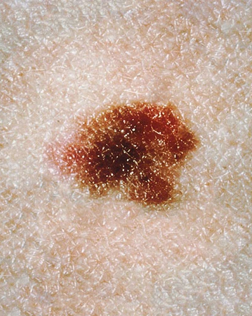
You might notice the following features, often remembered by the ABCDEs of moles:
- Asymmetry: One half of the mole does not match the other half.
- Border: The edges are irregular, scalloped, or poorly defined.
- Color: The color is varied from one area to another; shades of tan, brown, black, and sometimes red, pink, or white.
- Diameter: Usually larger than common moles (often larger than 6mm, about the size of a pencil eraser), but they can be smaller.
- Evolving: The mole looks different from others on your skin or is changing in size, shape, or color over weeks or months.
- Itching or tenderness in the mole, though this is less common.
- A flat component, often with a raised, "pebbly" or "cobblestone" surface.
- They can appear anywhere on the body, but are most common on sun-exposed areas like the back, chest, and legs.
Diagnosis
A dysplastic nevus is typically diagnosed by a healthcare professional, often a dermatologist, during a skin examination. They will look at the mole's size, shape, color, and texture.
Your doctor may use a special magnifying tool called a dermatoscope to see the mole's pattern more clearly. If a mole looks suspicious, a skin biopsy may be performed. This involves removing a small sample of the mole (or the entire mole) to be examined under a microscope by a pathologist to confirm the diagnosis and rule out melanoma.
Management & Treatment
Finding out you have an atypical mole can be worrying, but it’s important to remember that having one doesn’t automatically mean you have skin cancer. The main goal is to monitor these moles closely and remove any that look suspicious to prevent them from potentially developing into melanoma.
Most atypical moles do not need treatment and are monitored instead. Your dermatologist will guide you on the best approach, which depends on factors like the mole's level of atypia (how abnormal the cells look), your number of moles, and your personal and family medical history.
Observation and Self-Care
- Regular Skin Checks: The most important step is to keep an eye on your skin. We recommend monthly self-exams to check for any new moles or changes in existing ones. Use the ABCDEs of melanoma as your guide (Asymmetry, Border, Color, Diameter, Evolving).
- Professional Monitoring: Your dermatologist will likely want to see you for regular professional skin exams. This could be every 6 to 12 months, or more frequently if you have many atypical moles or other risk factors. They may take photos of your moles to track them over time, a practice called "mole mapping."
- Sun Protection is Key: Protecting your skin from the sun is a critical part of managing atypical moles. UV exposure can increase your risk of developing melanoma. Make it a habit to use a broad-spectrum sunscreen with an SPF of 30 or higher, wear sun-protective clothing, and avoid tanning beds completely.
Medical Procedures
If a mole is particularly concerning or if a biopsy shows moderate to severe changes, your dermatologist will recommend removing it.
- Surgical Excision: This is the most common procedure. The dermatologist will numb the area with a local anesthetic and then surgically cut out the entire mole along with a small border of normal-looking skin around it. This border, or "margin," helps ensure all the atypical cells are removed. The removed tissue is then sent to a lab to be examined.
- Stitches and Healing: You may need a few stitches after the removal. Your doctor will give you clear instructions on how to care for the wound to prevent infection. The area typically heals within one to two weeks.
Important Warning: Please never attempt to remove a mole at home. This can be dangerous, lead to infection and scarring, and—most critically—if the mole is cancerous, it can leave cancer cells behind that could spread.
When to Follow Up
Deciding on the right path—whether to watch a mole or remove it—is a decision you and your dermatologist will make together. If a mole is removed, the lab results will confirm if any further steps are needed. If you are monitoring your moles and notice any changes, it is crucial to contact your dermatologist right away. Your watchfulness is the best tool for catching any potential problems early.
Duration & Outlook
Dysplastic nevi are generally stable and can remain unchanged for many years; they are considered chronic in that they don't usually disappear on their own. While most dysplastic nevi will not turn into melanoma, having them does indicate an increased risk of developing melanoma at some point in life, either within an existing dysplastic nevus or as a new spot elsewhere on the skin.
Regular self-skin exams and professional skin checks are very important for monitoring. Warning signs for complications include any changes in an existing mole or the appearance of a new, suspicious-looking mole, especially if it follows the ABCDE criteria.
Prevention
While you can't always prevent dysplastic nevi, especially if you have a genetic predisposition, you can take steps to reduce your risk and catch changes early:
- Practice sun safety: Limit sun exposure, especially during peak hours (10 a.m. to 4 p.m.). Wear protective clothing, wide-brimmed hats, and sunglasses. Use broad-spectrum sunscreen with an SPF of 30 or higher daily, even on cloudy days.
- Avoid tanning beds and sunlamps - entirely, as these significantly increase your risk.
- Perform regular self-skin exams - (monthly) to become familiar with your moles and identify any new or changing spots.
- Schedule regular professional skin examinations - with a dermatologist, especially if you have many moles, a family history of melanoma, or previous dysplastic nevi. The frequency will be recommended by your doctor.
Causes & Triggers
The exact cause of dysplastic nevi isn't fully understood, but it's believed to be a combination of:
- Genetic factors: A tendency to develop atypical moles can run in families. Certain inherited gene mutations can increase the risk.
- Ultraviolet (UV) radiation exposure: Exposure to UV light from the sun or artificial sources like tanning beds is a major trigger and risk factor.
Who is most likely to develop it?
- Individuals with a personal or family history of dysplastic nevi or melanoma.
- People with many common moles (typically more than 50).
- Those with fair skin, light-colored eyes, and red or blonde hair who sunburn easily.
- People with a history of excessive sun exposure or severe sunburns, especially during childhood.
- Individuals with weakened immune systems.
When to see a doctor
It's important to see a healthcare professional, preferably a dermatologist, if you notice:
- Any new mole - that looks unusual or different from your other moles.
- Any change in an existing mole’s - size, shape, color, or elevation.
- A mole that develops symptoms like itching, tenderness, pain, bleeding, or crusting.
- A mole that looks like an "ugly duckling" – one that stands out or looks different from all other moles on your body.
Remember, early detection is key. While it can be worrying to notice a change, seeing a doctor promptly allows for accurate diagnosis and peace of mind or early intervention if needed.
Frequently Asked Questions (FAQs):
- Is a dysplastic nevus a type of cancer? No, a dysplastic nevus is not cancer. It is considered a benign (non-cancerous) mole, but it can be a marker for increased risk of developing melanoma, a serious skin cancer. Some dysplastic nevi can, rarely, develop into melanoma over time.
- Do all dysplastic nevi need to be removed? Not necessarily. Your dermatologist will assess each mole. If a dysplastic nevus shows highly atypical features, is changing, or if there's a strong personal or family history of melanoma, removal might be recommended for biopsy and to prevent potential progression. Many stable dysplastic nevi are simply monitored.
- If I have one dysplastic nevus, will I get more? It's possible. Some people are prone to developing multiple dysplastic nevi. Regular skin checks are important to monitor existing moles and identify any new ones.
- Can a dysplastic nevus turn into melanoma? Yes, although the risk for any single dysplastic nevus is low, they do have a higher potential to transform into melanoma compared to common moles. This is why monitoring is so important.
- How often should I get my skin checked if I have dysplastic nevi? Your dermatologist will recommend a schedule based on your individual risk factors, such as the number and appearance of your moles, your personal history, and your family history of skin cancer. This could range from every few months to annually.
This information is for educational purposes and should not replace advice from a healthcare professional. If you have concerns about a mole, please consult a doctor or dermatologist.